
IB Syllabus Focus:
'Examine the structure of prokaryotic cells, including components like cell wall, plasma membrane, cytoplasm, naked DNA, and 70S ribosomes.'
Prokaryotic cells, representing the simplest cellular organisms, are found throughout our world, from our bodies to extreme environments like thermal vents and acidic lakes. Their unique structure sets them apart from more complex eukaryotic cells. The domains of life that are classified as prokaryotes include Bacteria and Archaea. Not having a nucleus or membrane-bound organelles, they boast a straightforward design, yet play monumental roles in ecology, evolution, and human health.
Key Components of Prokaryotic Cells
Peptidoglycan: A polymer consisting of sugars and amino acids that forms a strong, mesh-like layer outside the plasma membrane of most bacteria, providing structural strength and shape.
Cell Wall
Function: The cell wall gives the cell its shape, offers protection, and combats osmotic pressures.
Composition:
Bacteria predominantly have a peptidoglycan-based cell wall.
Archaea, on the other hand, lack peptidoglycan, instead possessing walls made of various other polysaccharides and proteins.
Gram Staining: This technique divides bacteria into two groups:
Gram-positive bacteria, which have a thick peptidoglycan layer, appear purple.
Gram-negative bacteria have a thinner layer and an additional outer lipid membrane, staining pink or red.
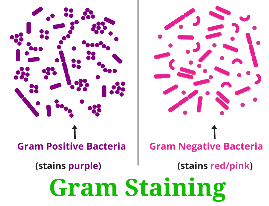
Image courtesy of RBR Life Science
Plasma Membrane
Phospholipid Bilayer: A double layer of phospholipid molecules with hydrophilic heads facing outward and hydrophobic tails facing inward, forming the fundamental structure of cell membranes.
Function: This semipermeable barrier manages the flow of molecules in and out of the cell, ensuring that essential molecules like nutrients enter and waste products exit.
Structure: The plasma membrane is made up of a double layer of phospholipids, with protein molecules interspersed.
Fluid Mosaic Model: This concept emphasises the dynamic, fluid nature of the membrane. The proteins and lipids can move laterally, giving the membrane its flexibility.
Cytoplasm
Function: Serving as the site of most cellular activities, the cytoplasm houses all the cell's internal components (except DNA).
Composition: A thick, jelly-like substance, it is primarily water but also contains enzymes, salts, and organic molecules.
Role in Metabolism: It's the arena for various metabolic reactions, driven by enzymes that accelerate chemical reactions vital for the cell's existence.
Enzyme: A biological catalyst, usually a protein, that accelerates the rate of specific chemical reactions without being consumed in the process.
Naked DNA
Function: It provides the instructions necessary for building and maintaining the cell.
Nucleoid: The irregularly shaped region within a prokaryotic cell where the naked, circular DNA molecule is located, lacking a surrounding membrane.
Structure: In prokaryotes, the DNA is not enclosed within a nuclear envelope, making it "naked." It's typically circular, lacking the complex proteins eukaryotes possess.
Location: Though not contained within a true nucleus, prokaryotic DNA is found within a defined region called the nucleoid.
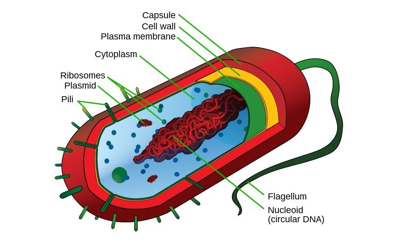
Image courtesy of Mariana Ruiz Villarreal, LadyofHats
70S Ribosomes
70S Ribosomes: These are the cellular machines responsible for protein synthesis.
Structure: A ribosome is composed of RNA and proteins and, in prokaryotes, is made of a 50S (large) and a 30S (small) subunit.
Significance: The distinction in size and structure between prokaryotic 70S ribosomes and eukaryotic 80S ribosomes is crucial. Some antibiotics target bacterial ribosomes, harming the bacteria without affecting the host.
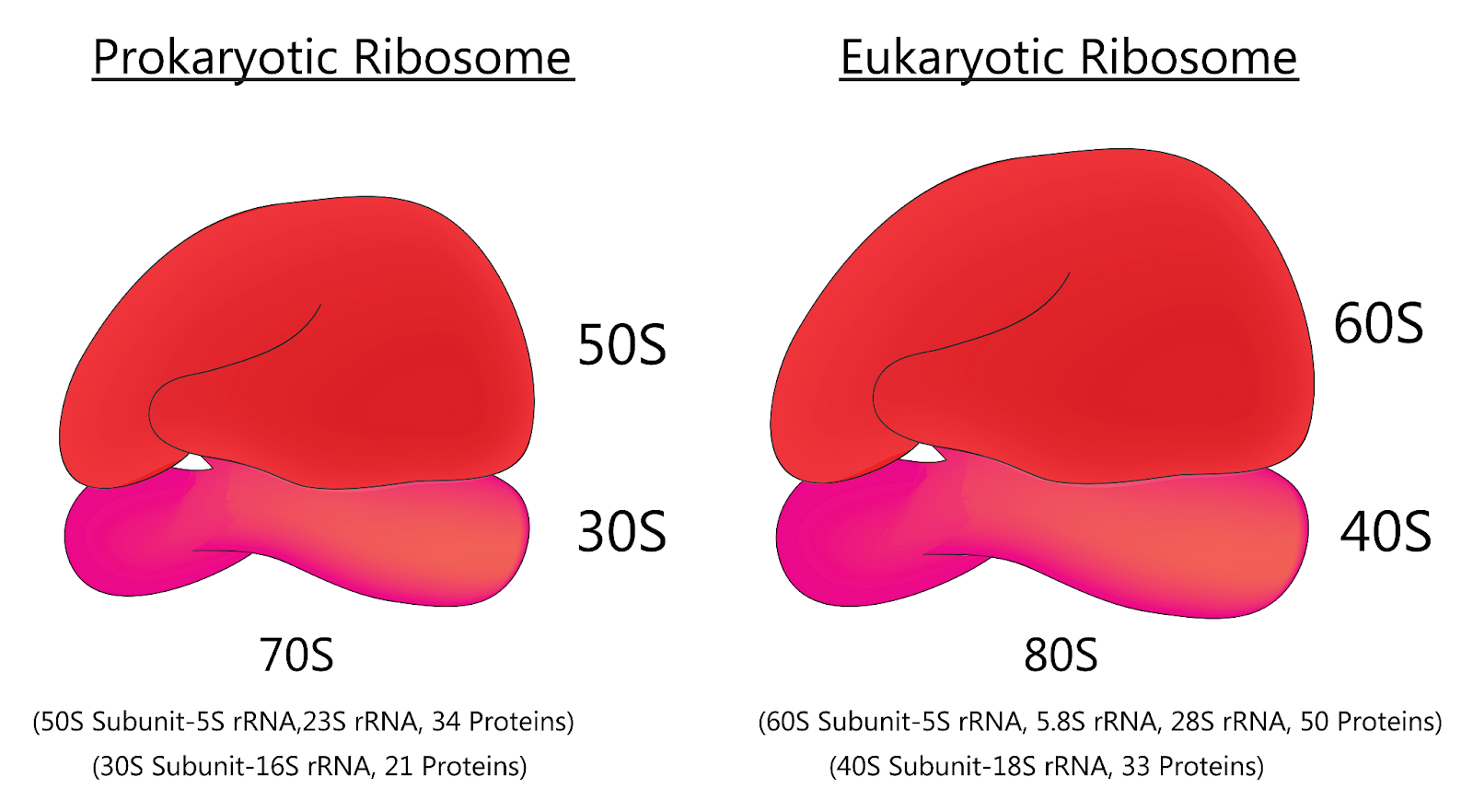
Image courtesy of KKT Madhusanka
Additional Prokaryotic Structures
Flagella and Pili
Conjugation: The process by which one prokaryotic cell transfers genetic material to another through direct contact, often using a pilus.
Function: Facilitate movement and attachment.
Flagella: Long, whip-like structures propelling the cell forward.
Pili: Shorter structures aiding in attachment to surfaces or other cells and playing a role in DNA transfer during conjugation.
Storage Granules
Function: Store reserve compounds, such as phosphates or energy-rich polysaccharides, for times when the cell might face nutrient scarcity.
Endospores
Function: These are durable, dormant structures formed during adverse conditions, ensuring the bacterium's survival. When conditions become favourable, the endospore can revert to a vegetative state.
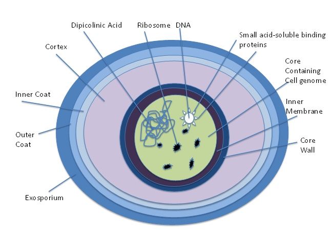
A detailed structure of endospore.
Image courtesy of Alayna5231
Differences from Eukaryotic Cells
Beyond the aforementioned distinctions:
Complexity: Prokaryotic cells are much simpler, lacking the membrane-bound organelles found in eukaryotic cells.
Reproduction: Prokaryotes reproduce asexually through binary fission, a simpler and quicker process than eukaryotic cell division.
Binary Fission: A method of asexual reproduction in prokaryotes where a single cell divides into two genetically identical daughter cells after DNA replication.
Relevance of Prokaryotic Cells
Understanding prokaryotes isn't just academic; it has real-world implications:
Biotechnology: Prokaryotes are used in biotechnology for producing medicines, bioremediation, and food production.
Health and Medicine: Knowledge of bacterial structure aids in creating treatments for bacterial infections.
Ecology: Prokaryotes are foundational to many ecosystems, contributing to nutrient cycling and forming symbiotic relationships with other organisms.
FAQ
Many prokaryotic cells are motile, and they achieve movement primarily using structures like flagella and, to a lesser extent, pili. Flagella are long, whip-like structures that extend from the cell's surface. These structures rotate in a propeller-like manner, allowing the cell to move towards or away from stimuli, a behaviour termed as chemotaxis. Pili, while primarily known for attachment and DNA transfer, can also aid in a type of movement called 'twitching motility' in some bacteria. The combination of these structures ensures that prokaryotic cells can navigate their environment effectively, responding to favourable or adverse conditions.
Prokaryotic cells, particularly certain bacteria, have developed a unique survival mechanism to withstand unfavourable conditions by forming endospores. Endospores are tough, dormant structures that encapsulate the cell's genetic material and some cytoplasm in a protective shell. This shell is resistant to extreme temperatures, radiation, chemical disinfectants, and dehydration. When environmental conditions become favourable again, these endospores can germinate and revert back to their vegetative state, ensuring the bacteria's survival and propagation. This ability is why certain bacteria, like Bacillus anthracis (which causes anthrax), are particularly hardy and challenging to eradicate.
Yes, there are other cellular structures in prokaryotes not covered in the primary syllabus. One notable example is the capsule, a sticky layer surrounding some bacterial cells, primarily composed of polysaccharides. It aids bacteria in adhering to surfaces, evading host immune responses, and preventing dehydration. Another structure is the plasmid, which are small, circular DNA fragments separate from the main bacterial chromosome. Plasmids often carry genes that confer specific advantages to bacteria, like antibiotic resistance. These structures, while not exhaustive in the study notes, further illustrate the complexity and adaptability of prokaryotic cells in various environments.
The nucleoid is a distinct region within prokaryotic cells where the circular DNA molecule is found. Unlike eukaryotic cells, prokaryotes lack a defined nucleus with a nuclear envelope. Instead, their "naked" DNA, which isn't associated with histones or packed into chromosomes, resides within the nucleoid region. This area isn't separated from the cytoplasm by any membrane, allowing direct interaction between the cell's genetic material and its metabolic machinery. The nucleoid ensures that the cell's DNA remains organised, protected, and accessible for crucial processes like transcription and replication.
Gram staining is a technique used to categorise bacterial species based on the structure of their cell walls. Gram-positive bacteria have a thick peptidoglycan layer in their cell walls which retains the crystal violet stain, thus appearing purple under a microscope. Gram-negative bacteria, on the other hand, have a thinner peptidoglycan layer and an outer lipid membrane. This outer membrane prevents the crystal violet stain from binding effectively, and so, after a counterstain with safranin, they appear pink or red. The distinction is essential as it provides insights into the bacterial cell's vulnerability to antibiotics and its potential pathogenicity.
Practice Questions
The plasma membrane in prokaryotic cells is a crucial semi-permeable barrier responsible for regulating the entrance and exit of molecules within the cell. It is composed of a bilayer of phospholipids interspersed with protein molecules. This configuration ensures that essential nutrients and ions can be taken up by the cell, while waste products are effectively removed. Furthermore, the plasma membrane follows the fluid mosaic model, where proteins and lipids can move laterally, offering flexibility to the membrane. This dynamic nature enables the membrane to facilitate various functions, such as transport, enzyme activity, and signal reception, supporting the cell's metabolic and communication processes.
Prokaryotic cells contain 70S ribosomes, made up of a 50S large subunit and a 30S small subunit, whereas eukaryotic cells possess 80S ribosomes with a 60S large subunit and a 40S small subunit. This distinction in structure and size is crucial from a medical standpoint. Some antibiotics specifically target the bacterial 70S ribosomes, inhibiting protein synthesis in the bacteria without affecting the eukaryotic 80S ribosomes present in the host organism. As a result, these antibiotics can effectively combat bacterial infections without harming the patient's cells. This selective targeting is a key principle in antibiotic design and usage.
