
IB Syllabus Focus:
'Explore advancements in microscopy techniques, such as electron microscopy, freeze fracture, cryogenic electron microscopy, fluorescent stains, and immunofluorescence.'
The journey of understanding the minuscule world of cells has been significantly propelled by advancements in microscopy techniques. These methods have revolutionised how we visualise and analyse cellular structures.
Electron Microscopy (EM)
Since its inception in the 1930s, electron microscopes have provided detailed glimpses into the cellular universe, far beyond the capabilities of traditional light microscopes.
Principle: Electron microscopes operate using beams of electrons instead of visible light. The behaviour of these electrons, when passed through or bounced off a specimen, is used to generate images.
Resolution: The ability of a microscope to distinguish two separate points as distinct objects; higher resolution reveals finer structural details.
Resolution: EM offers a remarkable resolution, capable of distinguishing structures approximately 0.1 nm apart.Transmission Electron Microscope (TEM)
Transmission Electron Microscope (TEM): A type of electron microscope that passes a beam of electrons through a very thin specimen to form highly detailed images of internal cellular structures.
Function: The TEM directs electrons through an ultra-thin specimen. As these electrons interact with cellular components, different structures absorb varying amounts, producing diverse image contrasts.
Image Representation: Structures appear darker or lighter based on how many electrons they absorb or transmit. This variation helps in visualising various cellular compartments.
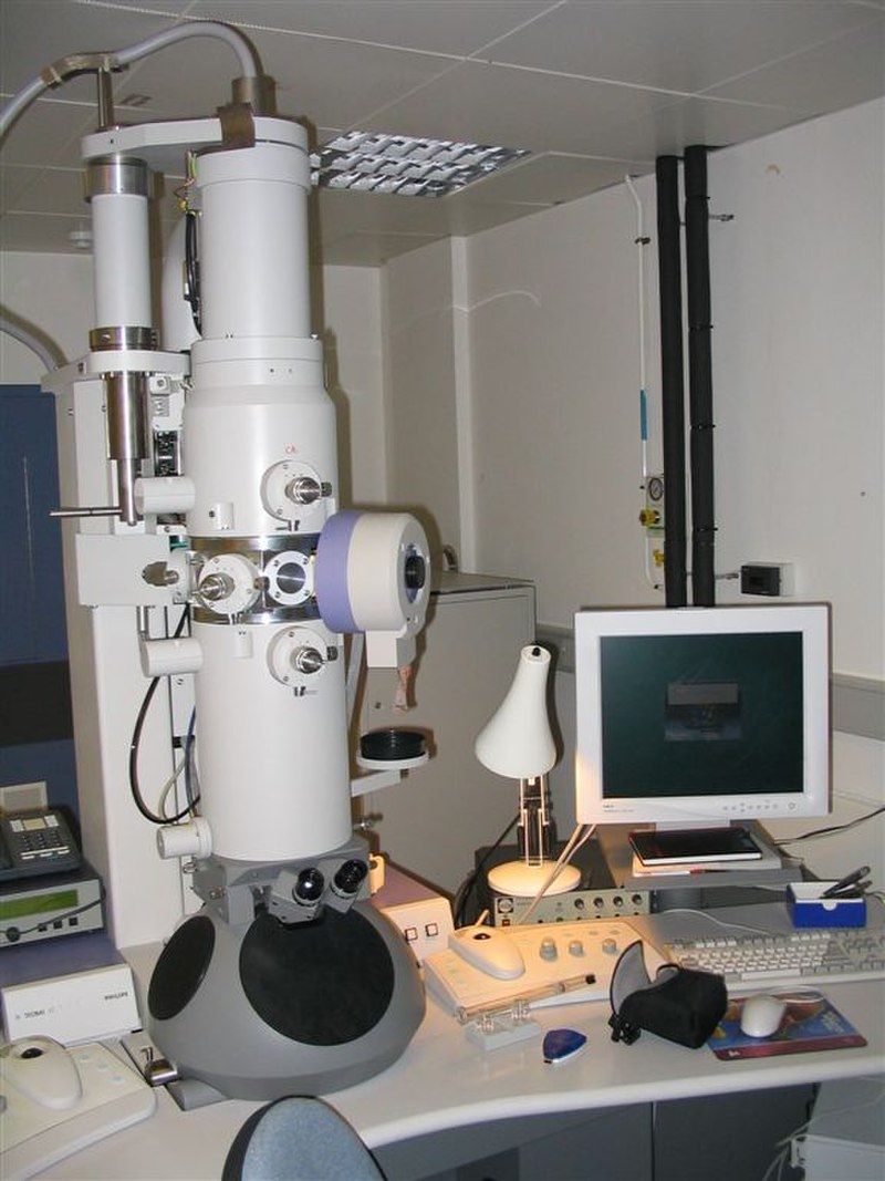
Transmission Electron Microscope
Image courtesy of David J Morgan
Scanning Electron Microscope (SEM)
Scanning Electron Microscope (SEM): An electron microscope that scans a focused beam of electrons across the surface of a specimen to produce detailed three-dimensional images of its surface topography.
Function: Instead of passing through the sample, electrons in SEM are bounced off the surface, rendering a three-dimensional representation.
Image Representation: The resulting images are intricate and detailed, offering a 3D view of the specimen's surface topology.
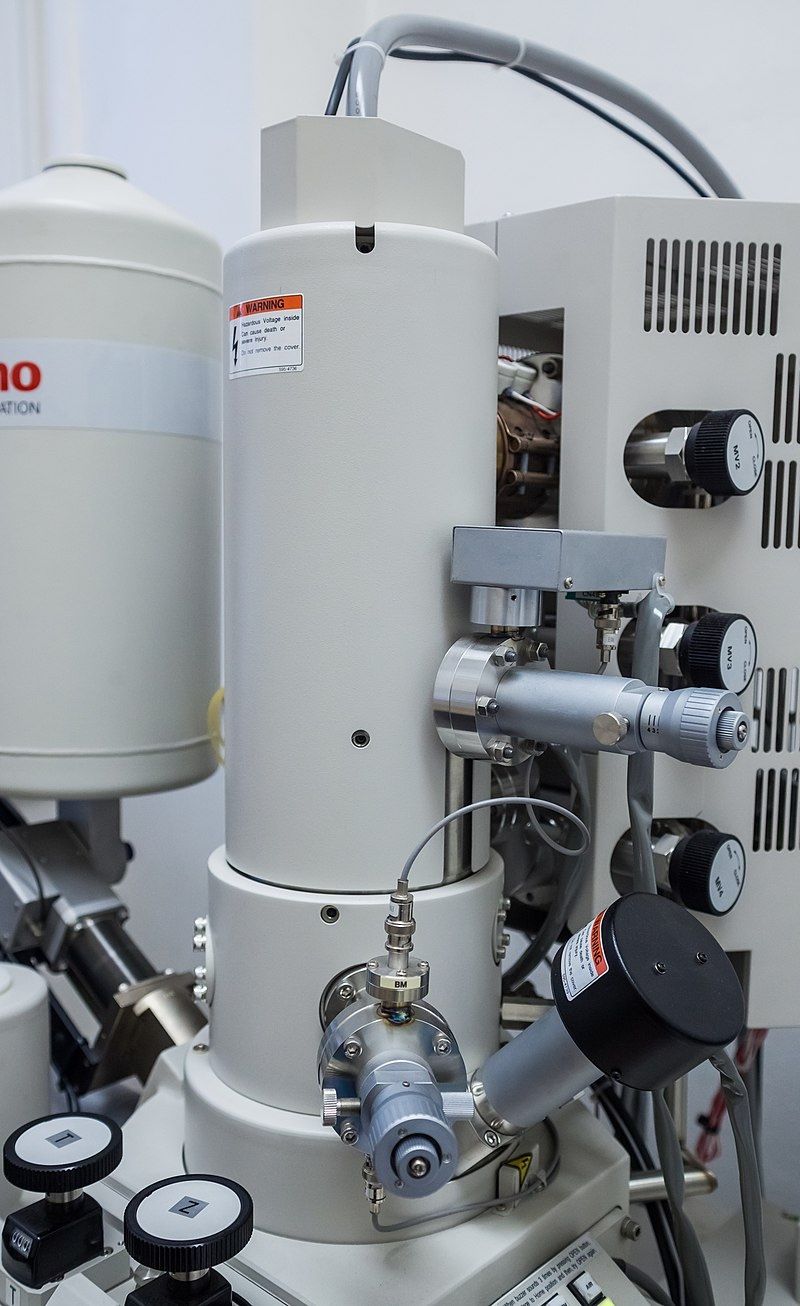
Scanning Electron Microscope
Image courtesy of Tadeáš Bednarz
Freeze Fracture
Freeze Fracture: A preparation technique in electron microscopy in which rapidly frozen cells are cracked open to reveal internal membrane structures for high-resolution imaging.
This technique is tailor-made for the in-depth study of membrane structures.
Process:
Cells or tissues undergo rapid freezing, solidifying all cellular components.
The frozen specimen is then fractured, usually splitting along lipid bilayer lines.
The exposed fractured surface is thinly coated with metal, producing a replica.
This replica is observed under the electron microscope.
Advantage: The method provides a deep insight into the interior landscapes of the cell membrane, including the positioning of embedded proteins.
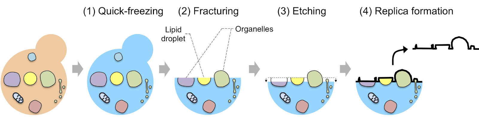
Image courtesy of Bio-protocol
Cryogenic Electron Microscopy (Cryo-EM)
Cryogenic Electron Microscopy (Cryo-EM): An imaging method where biological specimens are rapidly frozen to preserve their natural state, allowing high-resolution visualisation of molecular complexes without chemical fixation or staining.
A more recent advancement, Cryo-EM, combines the power of electron microscopy with cryogenic techniques.
Principle: Specimens are rapidly frozen, often in liquid ethane, preserving their native, hydrated state.
Advantages:
The method eliminates the need for staining or extensive sample preparation, which can sometimes alter cellular structures.
It offers exceptionally high-resolution images, ideal for visualising large molecular assemblies or cellular machinery.
Cryo-EM has been pivotal in the realm of structural biology, enabling detailed views of protein structures, virus particles, and more.
Fluorescent Stains
The advent of fluorescent stains has transformed cellular observations under the microscope.
Fluorescence: The emission of light by a substance that has absorbed light or electromagnetic radiation, commonly used to label specific cellular structures in microscopy.
Function: These are special molecules that, when exposed to specific wavelengths of light, emit light of a different wavelength. This phenomenon is harnessed to visualise specific cellular components.
Examples:
DAPI: Primarily binds to DNA, thus illuminating the cell nucleus under a fluorescence microscope.
FITC and Rhodamine: These are often used in combination to stain different cellular structures or molecules, enabling multi-colour imaging of specimens.
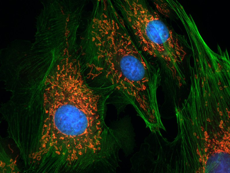
Endothelial Cells Fluorescent Image. Blue in the centre is nucleus stained with DAPI. Actin is shown in green and mitochondria in red.
Image courtesy of Erin Rod
Immunofluorescence
An elegant union of immunology and fluorescence microscopy.
Antibody: A Y-shaped protein produced by the immune system that specifically recognises and binds to an antigen, allowing targeted visualisation when tagged with fluorescent dyes in immunofluorescence techniques.
Process:
Specific antibodies, which can bind to a particular cellular component or protein, are produced.
These antibodies are then conjugated with fluorescent dyes.
When added to a specimen, these fluorescently-tagged antibodies bind to their target structures.
Under a fluorescent microscope, these targets become visible as brightly lit structures.
Applications:
It's extensively used in cell biology and pathology to determine the distribution of proteins, glycoproteins, and other antigens in cells.
It helps in cell differentiation, as certain cell types express unique proteins. By targeting these proteins, specific cell types can be identified and visualised.
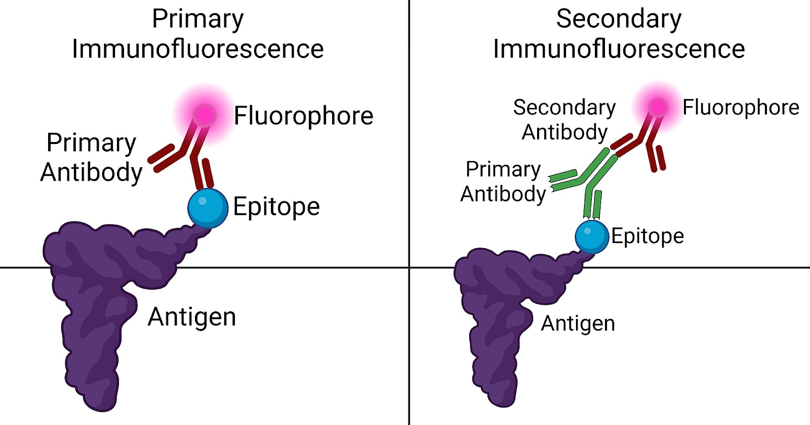
Image courtesy of Westhayl618
Ponder Over These Takeaways
Electron Microscopy is a cornerstone in modern cell biology, presenting a gateway to visualising the unseeable.
Freeze Fracture offers a peek into the intricate world of cell membranes and their resident proteins.
Cryo-EM is like capturing a moment in time, preserving specimens in their most natural state, and then unveiling their secrets at high resolution.
The luminous world of Fluorescent Stains and Immunofluorescence has revolutionised cellular imaging, adding colour and specificity to formerly invisible cellular realms.
Practice Questions
Cryogenic Electron Microscopy (Cryo-EM) is a transformative development in the field of microscopy. Unlike traditional electron microscopy, where samples often require extensive preparation and staining that might alter cellular structures, Cryo-EM involves rapidly freezing specimens, usually in liquid ethane. This swift freezing preserves the specimens in their natural, hydrated state. As such, there's no need for staining, and the method reduces potential structural alterations. Particularly for studying large molecular assemblies, Cryo-EM offers exceptional resolution, which is crucial. These assemblies, like protein complexes or virus particles, have intricate details, and Cryo-EM can capture these with unparalleled clarity without disturbing the native conformation of the molecules.
Transmission Electron Microscopy (TEM) and Scanning Electron Microscopy (SEM) are two advanced types of electron microscopes, but they operate on distinct principles. TEM works by directing electrons through an ultra-thin specimen. As these electrons interact with cellular components, structures appear darker or lighter based on the number of electrons they absorb or transmit. In contrast, SEM bounces electrons off the specimen's surface, generating a detailed three-dimensional representation of the surface. One advantage of TEM is its ability to provide intricate details of the internal structures of cells, while SEM excels in offering a three-dimensional view of the specimen's surface topology, which is invaluable for studying surface structures and features.
FAQ
Fluorescent stains, or fluorophores, are molecules that emit light of one colour (or wavelength) when they are illuminated by light of a different colour. Different fluorescent stains have affinities for specific cellular components or molecules. For instance, some stains bind selectively to DNA or RNA, some to proteins, lipids, or carbohydrates, and others might target specific cellular organelles like mitochondria or the Golgi apparatus. The specificity of these stains is often based on chemical interactions between the stain and the cellular component. By using a combination of stains that emit light at different wavelengths, scientists can visualise multiple structures or molecules simultaneously in a single cell.
Electron microscopes operate in a vacuum, which is necessary because air molecules would scatter the electron beam, reducing the resolution of the resulting image. This vacuum environment is not compatible with living specimens as they require water and air to survive. Moreover, the sample preparation for electron microscopy, which might involve dehydration, embedding in resin, or even coating with a thin metal layer, can alter or damage the cellular structures. Additionally, the high-energy electron beam itself can damage biological samples. For these reasons, living specimens cannot be viewed using electron microscopes; they must be fixed, dehydrated, and often sectioned before observation.
Immunofluorescence employs antibodies that are specific to a particular antigen or protein in the cell. These antibodies are tagged with a fluorescent dye. When introduced to a sample, these fluorescently-labelled antibodies bind only to their specific target. This high specificity is attributed to the unique interaction between antibodies and antigens – each antibody recognises and binds to a particular antigen. In contrast, conventional fluorescent staining might rely on more general interactions, such as a dye binding to all DNA in a cell or all proteins. As a result, immunofluorescence allows for the precise visualisation of specific proteins or structures within the cell, while conventional fluorescent staining might highlight a broader category of cellular components.
The freeze fracture technique capitalises on the unique property of the lipid bilayer of cell membranes. When a frozen specimen is fractured, it often breaks along the lines of the lipid bilayer because this is the region with the weakest bonds. This results in one leaflet of the lipid bilayer sticking to each fractured face. By then examining this fractured surface with an electron microscope, scientists can get an in-depth view of the interior of the lipid bilayer, including the positioning and distribution of embedded proteins. The method essentially offers a 'cross-sectional' view of the membrane, making it invaluable for understanding membrane architecture and protein distribution.
Resolution in microscopy refers to the smallest distance between two points that can still be distinguished as separate entities. It essentially determines the clarity and level of detail that can be observed in an image. A microscope with higher resolution will be able to discern smaller structures and details than one with lower resolution. In biological research, many cellular structures, such as organelles, protein complexes, and DNA strands, are incredibly small. To study and understand these structures' functions, it's crucial to visualise them clearly. A high-resolution microscope allows researchers to observe, analyse, and understand the intricacies of these tiny biological entities, thereby driving forward our understanding of cellular functions and processes.
