
AQA Specification focus:
'- Comprehensive analysis of polysaccharides: starch, glycogen, cellulose.
- Correlation of their structural attributes to functions in animal and plant cells.
- Biochemical testing methods for identification and analysis.'
Polysaccharides, complex and diverse carbohydrates, are pivotal in various biological processes. This comprehensive analysis explores starch, glycogen, and cellulose, focusing on their structures, roles in organisms, and biochemical identification methods.
1.3.3.1 Introduction to Polysaccharides
Polysaccharides are high-molecular-weight carbohydrates, formed by the condensation of many monosaccharide units. Their significance lies in their roles as energy storage and structural components in both animal and plant cells. This section will explore the structures and functions of the three primary polysaccharides: starch, glycogen, and cellulose.
1.3.3.2 Starch: Energy Storage in Plants
Starch is a polysaccharide found in many plants, primarily in their storage organs like seeds and tubers. It serves as an essential energy reserve.
Amylose
Structure: Composed of 200 to 20,000 α-glucose units, amylose forms a helical structure. This compact, linear chain is due to the α-1,4-glycosidic bonds linking the glucose units.
Function: Its helical structure allows for dense packing, making amylose an efficient form of energy storage. Enzymes can hydrolyse the α-1,4-glycosidic bonds to release glucose.
Amylopectin
Structure: Larger than amylose, amylopectin can contain up to two million α-glucose units. The structure is branched, with α-1,4-glycosidic bonds forming the linear sections and α-1,6-glycosidic bonds at the branching points.
Function: The branched structure facilitates more rapid hydrolysis by enzymes, making glucose readily available for plant metabolism.
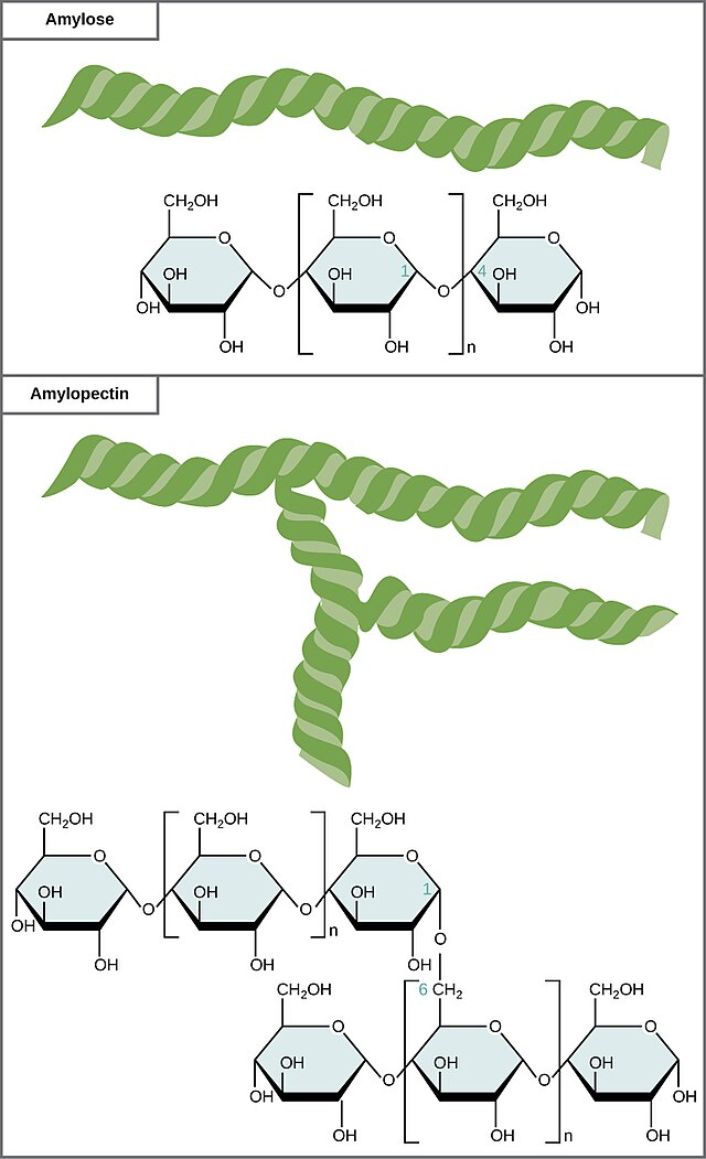
Image courtesy of CNX OpenStax
1.3.3.3 Glycogen: The Animal Starch
Glycogen is the primary storage form of glucose in animals, found mainly in the liver and muscle cells.
Structure: Highly branched, similar to amylopectin but more compact, with branches occurring every 8 to 10 glucose units.
Function: Glycogen's structure allows for rapid release of glucose when energy is required, crucial during physical activity and for maintaining blood glucose levels.
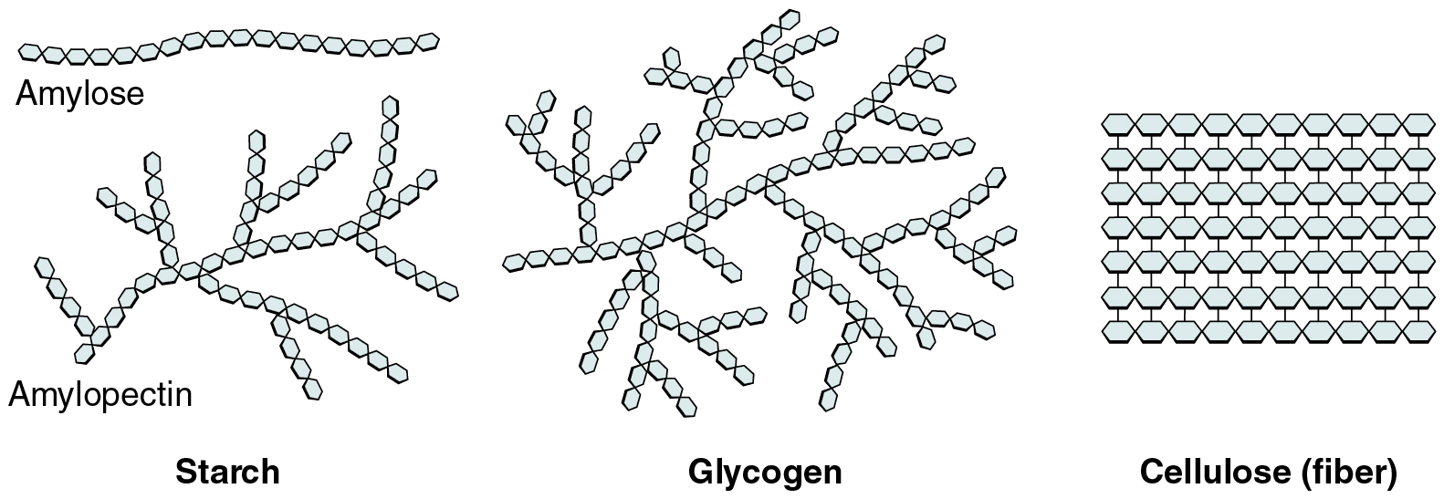
Image courtesy of OpenStax College
1.3.3.4 Cellulose: Plant Structure and Support
Cellulose is a fundamental component of plant cell walls and provides rigidity and strength.
Structure: Composed of β-glucose units linked by β-1,4-glycosidic bonds. The orientation of the glucose units results in straight, unbranched chains that form hydrogen bonds with each other, creating a rigid structure.
Properties: Its indigestibility in humans categorises it as a dietary fibre, contributing to digestive health.
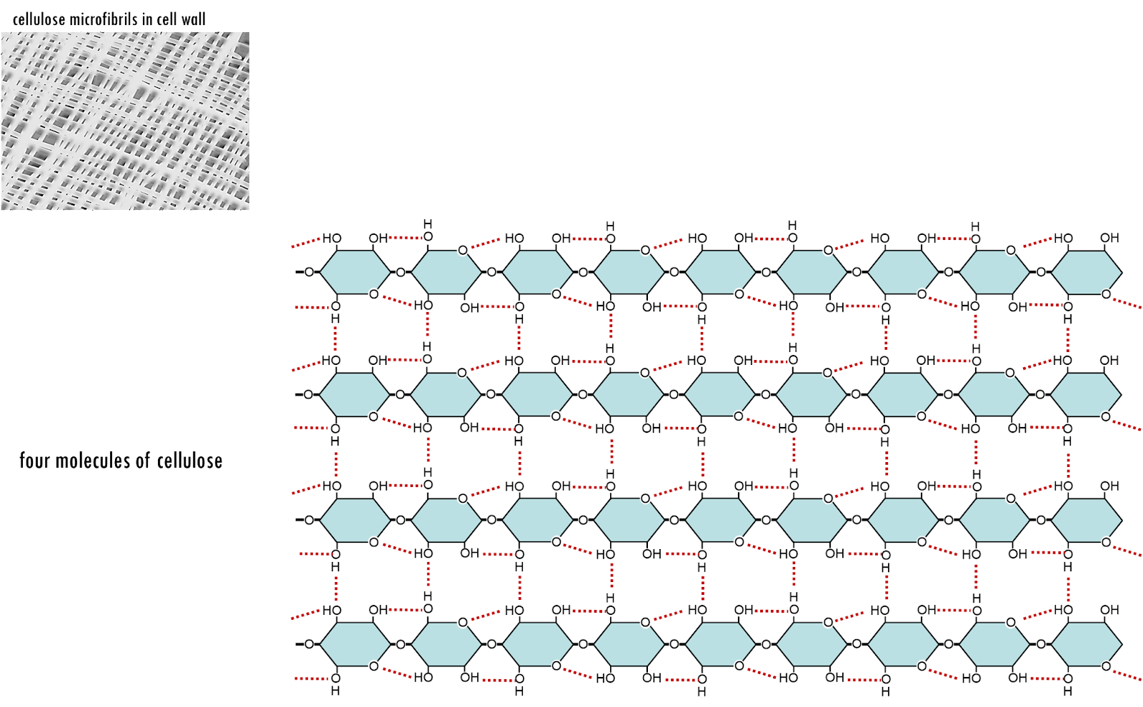
Image courtesy of Eunice Laurent
1.3.3.5 Functional Correlation of Polysaccharide Structures
The structural differences among starch, glycogen, and cellulose underpin their distinct functions:
Starch and Glycogen: Their structures, whether branched or helical, allow for compact storage and rapid mobilisation of energy.
Cellulose: The linear, rigid structure of cellulose provides mechanical support and protection to plant cells, crucial for maintaining plant architecture.
1.3.3.6 Biochemical Identification of Polysaccharides
Understanding the presence and type of polysaccharides in biological samples is essential for various biological studies and applications.
Iodine Test for Starch
Procedure: Iodine solution is added to a sample.
Observation: The interaction of iodine with the helical structure of amylose in starch results in a blue-black colouration, indicating the presence of starch.

Image courtesy of trinset
Benedict's Test for Non-Reducing Sugars
Procedure: Samples are hydrolysed with dilute acid, neutralised, and then Benedict's reagent is added.
Observation: A colour change from blue to green, yellow, orange, or brick-red indicates the presence of reducing sugars formed from the hydrolysis of non-reducing sugars.
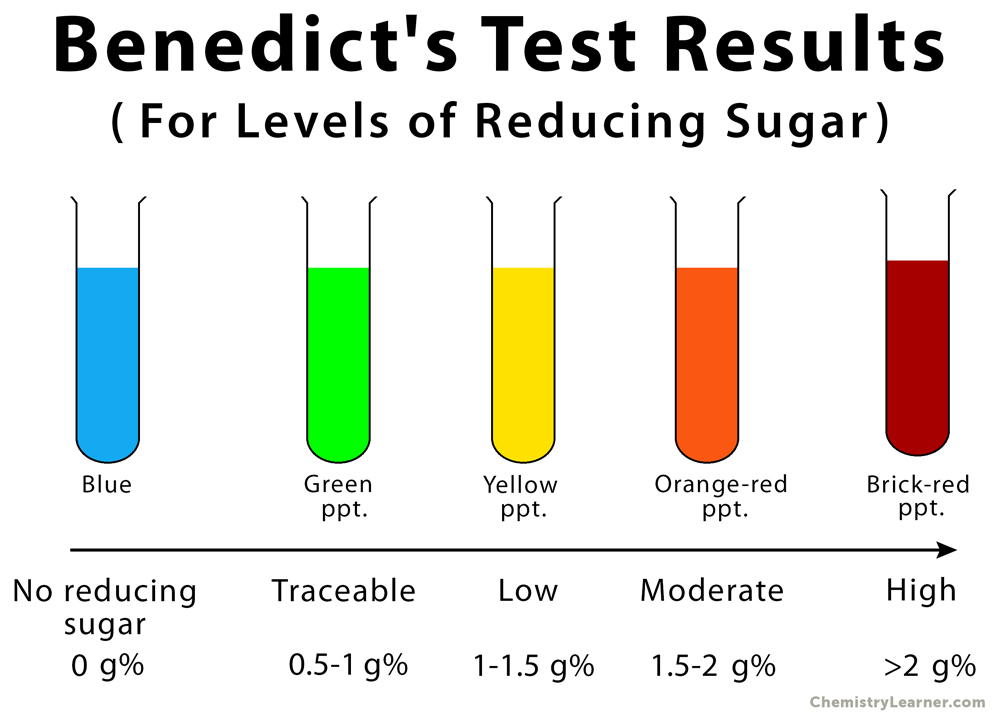
Image courtesy of Chemistry Learner
Biuret Test for Proteins
Importance: Conducted to ensure the absence of proteins in carbohydrate analysis.
Procedure: Biuret reagent (a mixture of copper sulfate and potassium hydroxide) is added to the sample.
Observation: A colour change from blue to purple indicates the presence of peptide bonds in proteins.
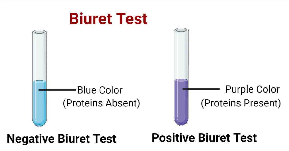
Image courtesy of Microbe Notes
1.3.3.7 The Biological Significance of Polysaccharides
Polysaccharides are not limited to energy storage and structural support; their roles extend into various biological processes:
Immune System: Specific polysaccharides on the surfaces of pathogens can trigger immune responses.
Dietary Fibre: Cellulose, an indigestible polysaccharide, aids in digestive health and can prevent certain gastrointestinal disorders.
Energy Metabolism: Glycogen plays a vital role in regulating blood sugar levels, crucial for energy balance and metabolic homeostasis.
Understanding the structure and function of polysaccharides provides insight into essential biological mechanisms. Their varied structures enable them to perform a wide range of functions critical for the survival and health of organisms. This knowledge is fundamental for students studying A-level biology, as it lays the foundation for more advanced concepts in biochemistry and cellular biology.
FAQ
The branching pattern in glycogen is more extensive compared to starch, particularly amylopectin, and this has significant implications for its function. In glycogen, branches occur every 8 to 10 glucose units, whereas in amylopectin, the branches are more spaced out. This dense branching in glycogen allows for more points of enzymatic attack, facilitating rapid release of glucose when energy is needed quickly, such as during physical exertion or in response to low blood sugar levels. This is crucial in animals, where immediate energy demands can be high. In contrast, starch, particularly amylose, has fewer branches, making it more suitable for long-term energy storage in plants. The structure of starch, with its mix of amylose and amylopectin, is optimised for gradual release of energy during germination and growth, in line with the energy requirements of plants.
Humans can digest starch but not cellulose due to the specific orientation of glucose molecules and the type of glycosidic bonds in these polysaccharides. Starch is composed of α-glucose units linked by α-1,4 and α-1,6 glycosidic bonds, which human digestive enzymes, such as amylase and glucosidase, can break down. In contrast, cellulose is made of β-glucose units linked by β-1,4-glycosidic bonds. Humans lack the enzyme cellulase, which is necessary to break these β-1,4 bonds. As a result, cellulose remains undigested, passing through the gastrointestinal tract as fibre. This difference in digestibility is due to evolutionary variations in the digestive systems of different organisms. Some animals, like ruminants, have symbiotic bacteria in their gut that produce cellulase, allowing them to digest cellulose.
Starch and glycogen, while both serving as energy storage molecules, have distinct roles in plants and animals due to their different physiological needs and lifestyles. In plants, starch is the primary energy reserve, stored in organs like seeds, roots, and tubers. It is utilised during periods of low photosynthetic activity, such as at night or during seed germination, providing a sustained energy source for growth and metabolic processes. The structure of starch, particularly its amylose component, is suited for long-term storage, releasing glucose gradually as needed. In contrast, glycogen is the main energy storage polysaccharide in animals. It is stored primarily in the liver and muscle tissues and is mobilised quickly in response to immediate energy needs, such as during sudden physical activity or to regulate blood glucose levels. The highly branched structure of glycogen allows for rapid glucose release, catering to the dynamic energy demands of animals.
Polysaccharides are identified and analysed in the laboratory using various biochemical tests and techniques. The most common tests include the iodine test for starch and Benedict's test for non-reducing sugars. The iodine test identifies starch by adding iodine solution to a sample, which forms a blue-black complex with amylose. Benedict's test, after hydrolysing polysaccharides into monosaccharides, detects reducing sugars through a colour change. Advanced techniques involve chromatography, such as gas-liquid chromatography (GLC) and high-performance liquid chromatography (HPLC), for separation and analysis of monosaccharide units. Spectroscopic methods like mass spectrometry and nuclear magnetic resonance (NMR) spectroscopy provide detailed structural information. These methods are crucial in research and industry, helping to understand the roles of polysaccharides in nutrition, medicine, and plant biology.
Cellulose, a major component of plant cell walls, exhibits a unique structure that is crucial for its function. It consists of long chains of β-glucose molecules linked by β-1,4-glycosidic bonds. These linear chains align parallel to each other, forming microfibrils through hydrogen bonding. This arrangement imparts high tensile strength and rigidity, essential for maintaining the structural integrity of plant cells. The rigidity of cellulose is vital for plant growth and upright posture, enabling plants to reach towards sunlight and maximise photosynthesis. Unlike starch and glycogen, cellulose does not serve as an energy reserve; its primary role is structural, providing support and protection. The indigestibility of cellulose in humans classifies it as dietary fibre, contributing to digestive health by promoting bowel movements and preventing constipation.
Practice Questions
Glycogen, with its highly branched structure comprising α-1,4 and α-1,6 glycosidic bonds, is excellently suited for its role as a quick-release energy store in animal cells. The branching significantly increases the surface area, allowing enzymes like glycogen phosphorylase to act simultaneously on many terminal glucose units. This facilitates rapid mobilisation of glucose when energy is required, particularly important during intense physical activity or between meals to maintain blood glucose levels. Additionally, the compact nature of glycogen's structure means it occupies minimal space within cells, allowing for efficient storage of glucose without significantly affecting cell volume.
The biochemical test for starch involves adding iodine solution to a sample. If starch is present, the iodine binds to the helical structure of amylose, a component of starch, causing a colour change to blue-black. This colour change is due to the formation of a polyiodide starch complex within the helical regions of amylose. The intensity of the blue-black colour can vary depending on the concentration of starch in the sample. This test is specific for starch as other carbohydrates do not form this coloured complex with iodine, allowing for the identification and qualitative analysis of starch in various biological and food samples.
