
IB Syllabus focus:
'- Principles of fMRI
- Differences between MRI and fMRI
- Applications and limitations'
Functional Magnetic Resonance Imaging (fMRI) is a neuroimaging technique allowing researchers to observe brain activity and determine which parts are involved in various mental processes. These notes will delve into the principles of fMRI, compare its aspects with MRI, and discuss its applications and limitations. To understand fMRI fully, it is beneficial to compare it with MRI Scans and PET Scans, which are also pivotal in neuroimaging.
Principles of fMRI
Basic Principle
fMRI operates on the principle of detecting changes in blood flow to different parts of the brain, which are indicative of neural activity. It leverages the blood-oxygen-level-dependent (BOLD) contrast imaging to visualise areas of the brain where neurons are especially active, showcasing higher oxygen levels. This process is closely linked to the localisation of function in the brain, an area extensively studied within neuroscience.
Working Mechanism
Neural Activity: When a brain region is more active, it consumes more oxygen.
Blood Flow: To compensate for the oxygen consumption, blood flow increases to the active area, bringing in oxygen-rich blood.
Magnetic Properties: The difference in magnetic properties of oxygen-rich and oxygen-poor blood is detected by fMRI. This phenomenon is further explained through synaptic transmission, which plays a significant role in neural communication.
Image Acquisition
Slices: fMRI acquires images in slices, which can be combined to create a 3D representation of the brain.
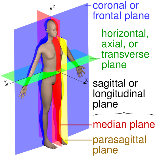
Anatomical imaging planes used in MRI: sagittal (median/parasagittal), coronal (frontal), and axial (transverse). Understanding these planes clarifies how slice-by-slice acquisition is assembled into a 3D brain volume in fMRI. Source.
Temporal Resolution: fMRI has high temporal resolution, capturing dynamic changes in the brain over time.
Differences between MRI and fMRI
Purpose
MRI (Magnetic Resonance Imaging):
Provides detailed images of the brain's structure.
Primarily used for diagnosing abnormalities in soft tissues.
fMRI:
Visualises brain activity.
Identifies regions involved in cognitive processes.
Imaging Contrast
MRI: Utilises differences in the water content of tissues to generate image contrast.
fMRI: Leverages the BOLD contrast to detect changes in blood oxygenation and flow, representing neural activity.
Timing
MRI: Typically focuses on static, structural images.
fMRI: Captures dynamic images to track brain activity over time.
Applications of fMRI
Cognitive Neuroscience
Brain Mapping: fMRI is pivotal in mapping the brain, enabling researchers to identify areas responsible for different functions and cognitive processes.
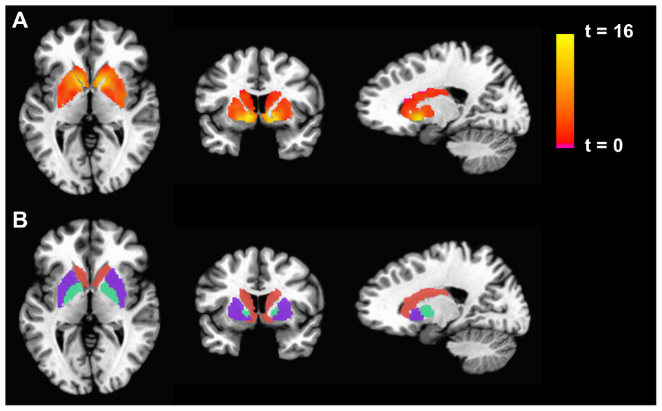
Functional activation maps (colored overlays) displayed on high-resolution structural MRI illustrate regions with significant BOLD signal change during task performance. This example contrasts basal ganglia activation in patient and control groups; while disease-specific, the visualization exemplifies how fMRI localizes function on anatomy. Source.
Mind-Brain Relationships: The technique aids in understanding the relationships between specific mental activities and the brain regions associated with them. The neuroplasticity page provides additional insight into how the brain adapts and changes in response to learning and experience, which is relevant to fMRI studies.
Clinical Psychology
Diagnosis: fMRI can help in the diagnosis of various neurological conditions and mental disorders by observing abnormal patterns of brain activity.
Treatment Efficacy: The technique is used to assess the efficacy of treatments and interventions by observing changes in brain activity patterns.
Research
Behavioral Studies: It’s extensively used in behavioral studies to analyse the neural substrates of human behaviour.
Comparative Analyses: Researchers utilise fMRI to conduct comparative analyses of brain activity under different conditions or stimuli.
Educational Insights
Learning Processes: fMRI sheds light on the neural mechanisms underlying learning and memory, offering insights into educational strategies and learning disabilities.
Limitations of fMRI
Temporal Resolution
Despite its high temporal resolution compared to other imaging techniques, fMRI might still miss rapid neural events due to the delay between neural activity and blood flow changes.
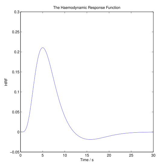
The canonical hemodynamic response function (HRF) shows the delayed increase and gradual return of the BOLD signal after brief neural activity. This delay reflects vascular dynamics rather than neurons firing directly, which is why very fast events can be missed in fMRI time series. Source.
Spatial Resolution
The spatial resolution of fMRI is lower than other techniques like EEG, making it challenging to precisely localise the origin of neural signals.
Cost and Accessibility
Expense: fMRI is expensive, limiting its availability and accessibility for research and clinical purposes.
Infrastructure: The infrastructure required for fMRI is extensive, and not all institutions have the necessary facilities.
Interpretation Challenges
Complexity: The complexity of the brain's network can make interpreting fMRI data difficult, with the risk of attributing mental functions to wrong brain regions.
Noise: The technique is sensitive to noise, which can be introduced by various sources such as the subject’s movements, affecting data accuracy.
Ethical Concerns
Privacy: The potential ability of fMRI to 'read minds' raises significant ethical concerns regarding privacy and consent. The discussion on ethical considerations in animal studies also highlights the broader ethical implications of using advanced neuroimaging techniques.
Overemphasis: There is a risk of overemphasis on brain-based explanations for behaviour, potentially neglecting socio-cultural factors and individual experiences.
Application Specificity
Movement Restriction: Subjects must remain still during scans, which might not be representative of brain activity during natural, real-world movements and experiences.
External Validity: The highly controlled environments necessary for fMRI studies may lack external validity, affecting the generalisability of the findings to real-world scenarios.
Practical Considerations
Safety
Non-invasive: fMRI is non-invasive, avoiding risks associated with invasive procedures.
Radiation-Free: It doesn’t use ionising radiation, making it safer for participants, especially compared to CT scans and X-rays.
Data Analysis
Advanced Processing: The data acquired needs advanced processing and analysis to interpret, requiring sophisticated software and expertise.
Individual Variability: The considerable variability in brain structure and function between individuals necessitates careful consideration during data interpretation.
In summary, the fMRI is a revolutionary tool in neuroscience and psychology, allowing researchers and clinicians to explore the human brain's mysteries. Its non-invasive nature and high temporal resolution make it a preferred choice for studying brain activity related to various cognitive processes, despite its limitations and challenges in data interpretation and ethical concerns.
Practice Questions
The principles of fMRI revolve around blood-oxygen-level-dependent (BOLD) contrast imaging, enabling the visualisation of active brain areas. When a brain region is active, it consumes more oxygen, leading to increased blood flow to supply oxygen-rich blood. fMRI detects changes in blood oxygenation and flow, reflecting neural activity. The technique captures images in slices, which are combined to create a 3D representation of the brain, offering insights into dynamic brain activities. The high temporal resolution of fMRI enables the tracking of rapid changes in neural activity, rendering it crucial for understanding brain functionality.
fMRI scans, despite their invaluable insights, have limitations like high costs, necessity for extensive infrastructure, and the challenge of accurately interpreting complex brain networks. The spatial resolution is also lower compared to other methods, causing difficulties in localising neural signals precisely. Ethically, fMRI poses concerns relating to privacy due to its potential to decipher thoughts, and there’s a risk of over-relying on brain-based explanations, neglecting socio-cultural and individual aspects. Additionally, the need for subjects to remain still and the highly controlled environments of studies might affect the external validity and generalisability of the findings.
FAQ
Indeed, there are differences in interpreting fMRI data for research versus clinical diagnosis. In research, the focus is often on understanding the general patterns of brain activity related to specific cognitive functions or tasks, requiring sophisticated statistical analyses and often involving group-level comparisons. This approach helps in developing theories about brain function and informing our understanding of neural underpinnings of behaviour. In contrast, clinical diagnosis primarily involves individual-level analyses, focusing on identifying abnormalities in brain activity or structure that are indicative of specific neurological conditions or mental disorders, aiming for accurate diagnosis and informing treatment plans.
Movement restrictions during fMRI scans can significantly impact study outcomes. Since participants must remain very still to avoid image blurring and distortion, the obtained results may not always reflect brain activity during natural, real-world movements and experiences. This limitation raises questions about the ecological validity of fMRI studies and the generalisability of the findings. The unnatural environment and the conscious effort to remain still might alter participants’ mental states and responses, potentially affecting the brain activity patterns that the study intends to measure and analyse.
fMRI significantly contributes to understanding neurological and psychological disorders by visualising aberrant brain activity patterns associated with various conditions. For example, it can highlight hypoactive or hyperactive regions in disorders like depression or schizophrenia, respectively. This visual representation aids clinicians in accurately diagnosing conditions, understanding their underlying neural substrates, and evaluating treatment efficacy. Moreover, by observing how brain activity changes in response to interventions, researchers can develop more effective treatment strategies and therapeutic approaches, paving the way for personalised medicine in neurology and psychology.
While fMRI is an advanced technique, it cannot detect all types of brain activity. It is primarily sensitive to changes in blood flow related to neural activity in the cerebral cortex and has difficulty detecting activity in subcortical regions. fMRI is limited by its reliance on the hemodynamic response, which has a slower onset than the actual neural activity, potentially missing brief neural events. Furthermore, fMRI mainly detects the input and processing activity of neurons in a region but may not accurately capture the output activity, leading to an incomplete picture of neural communication.
The quality of fMRI images is relatively high, providing a detailed view of brain activity by depicting blood flow. However, while fMRI offers superior spatial resolution compared to EEG and MEG, allowing for the identification of active brain regions with reasonable accuracy, it is not as precise as techniques like CT in revealing anatomical structures. Additionally, the quality of fMRI images is inherently linked to temporal resolution; the technique may fail to capture extremely rapid neural events due to the inherent delay in vascular response compared to neural activity.
