
Proteins, the workhorses of the cell, exhibit a remarkable diversity of functions, all of which are intricately tied to their complex structures. These structures can be broken down into four distinct levels – primary, secondary, tertiary, and quaternary – each contributing to the protein's unique attributes and capabilities.
Primary Structure
The primary structure of a protein is its linear sequence of amino acids, linked by peptide bonds. This sequence is determined by the gene encoding the protein and is fundamental in dictating the protein's final structure and function. Each of the 20 different amino acids imparts distinct chemical properties, influencing how the protein interacts with other molecules.
- Peptide Bonds: These are formed in a dehydration synthesis reaction, where a water molecule is released. The bond forms between the carboxyl group of one amino acid and the amino group of the next.
- Sequence Importance: A single alteration in the amino acid sequence (a mutation) can drastically change the protein's properties, as seen in disorders like sickle cell anaemia.
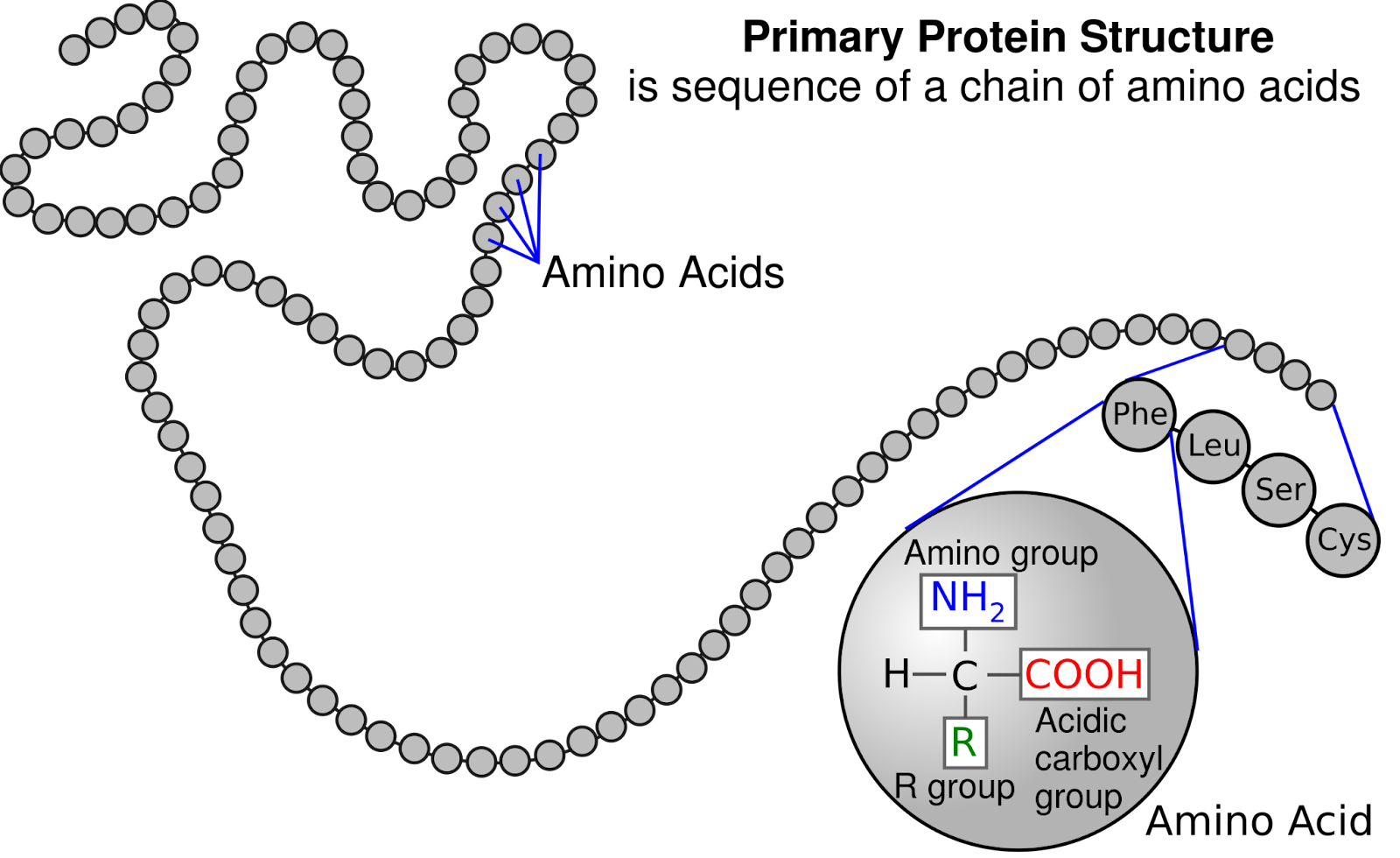
Image courtesy of National Human Genome Research Institute
Secondary Structure
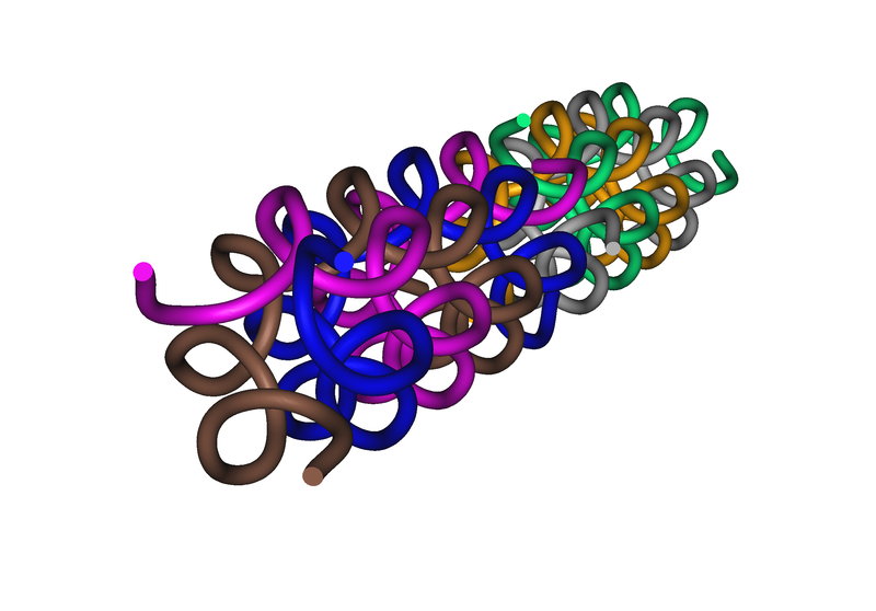
The secondary structure refers to the local folding or coiling of the polypeptide chain, primarily driven by hydrogen bonds. This level of structure includes alpha helices and beta-pleated sheets, both of which add stability and shape to the protein.
- Alpha Helices: These are right-handed coils where every backbone N-H group forms a hydrogen bond with the C=O group of the amino acid four residues earlier. This structure is common in fibrous proteins like keratin.
- Beta-Pleated Sheets: These sheets form when beta strands are held together by hydrogen bonds. Strands can be parallel (aligned in the same direction) or antiparallel (aligned in opposite directions).
- Importance of Hydrogen Bonds: These bonds are individually weak but collectively confer significant stability to the protein structure.
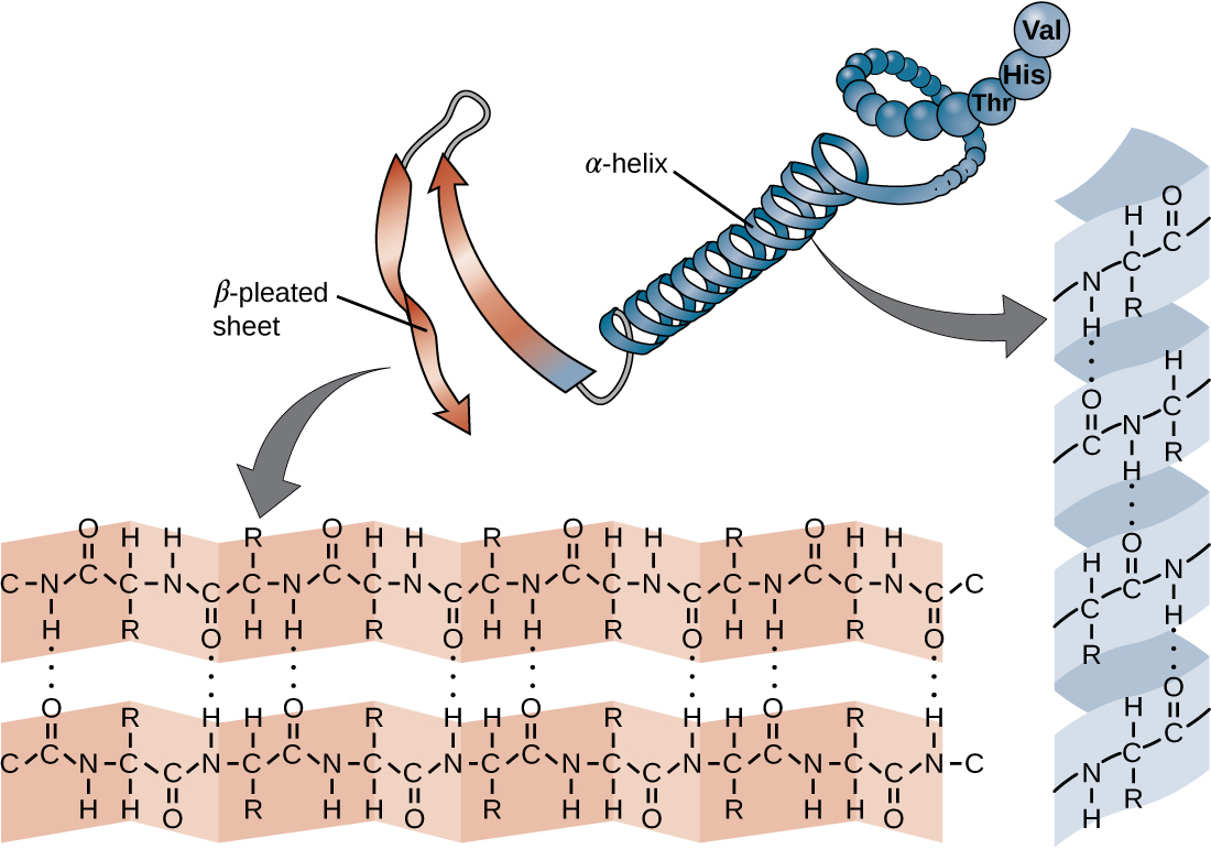
Image courtesy of CNX OpenStax
Tertiary Structure
Tertiary structure represents the complete three-dimensional structure of the protein. This level is defined by the interactions of the amino acid side chains, which include hydrophobic interactions, hydrogen bonds, ionic bonds, and disulfide bridges.
- Hydrophobic Interactions: These occur when non-polar side chains are buried in the protein core, away from water, driving the folding process.
- Ionic Bonds and Disulfide Bridges: Ionic bonds between oppositely charged side chains and covalent disulfide bridges between cysteine residues provide additional stability.
- Protein Domains: Tertiary structures often contain distinct functional regions known as domains, each with a specific role in the protein's function.
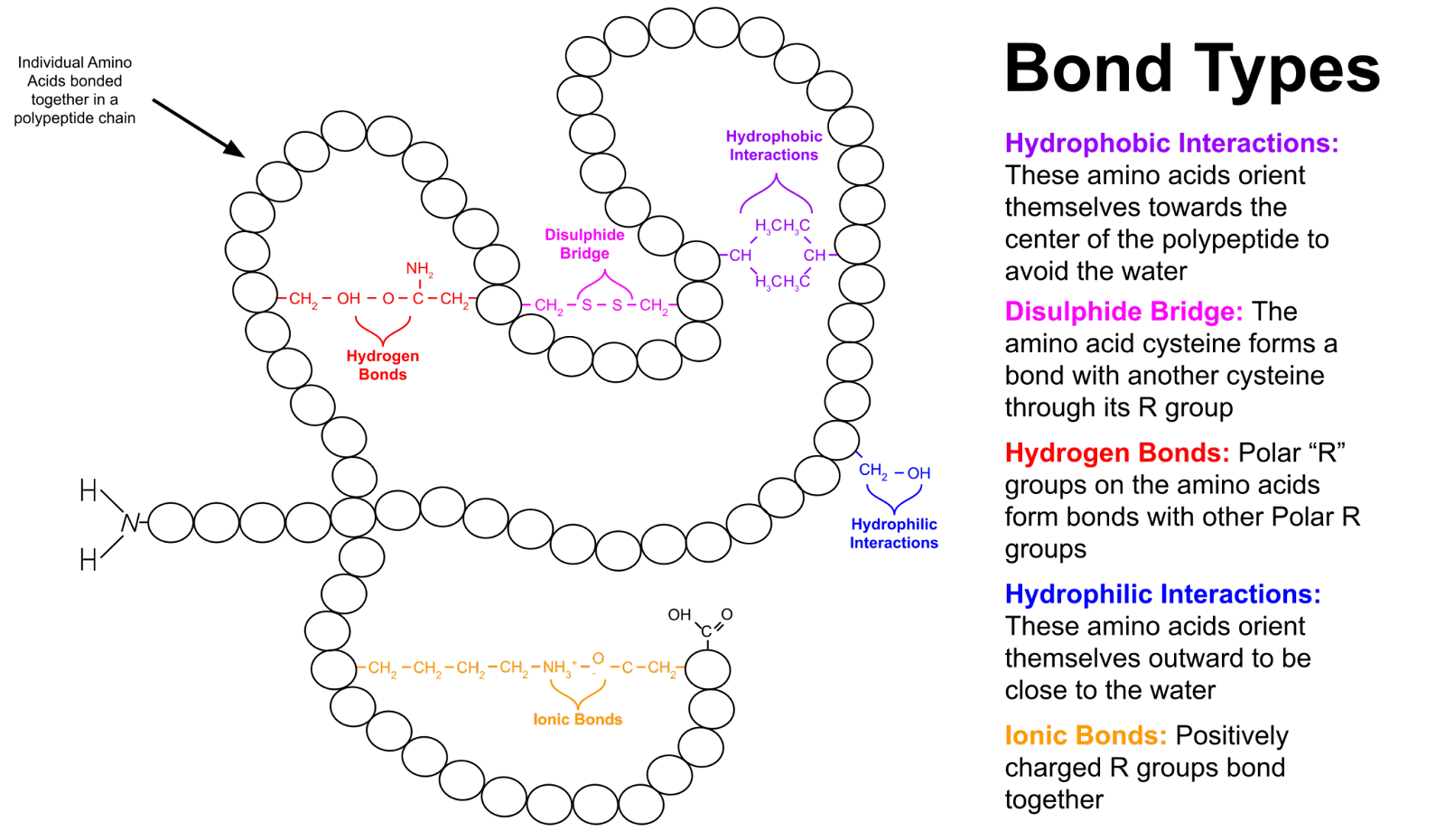
Image courtesy of WikiComTD
Quaternary Structure
Quaternary structure arises when multiple polypeptide chains (subunits) come together to form a functional protein complex. This level of structure is critical for proteins that operate as part of larger assemblies.
- Subunit Assembly: The arrangement of subunits is guided by the same types of interactions seen in tertiary structures.
- Examples and Significance: Hemoglobin, an oxygen-transport protein, is a classic example of a protein with quaternary structure, consisting of four subunits.
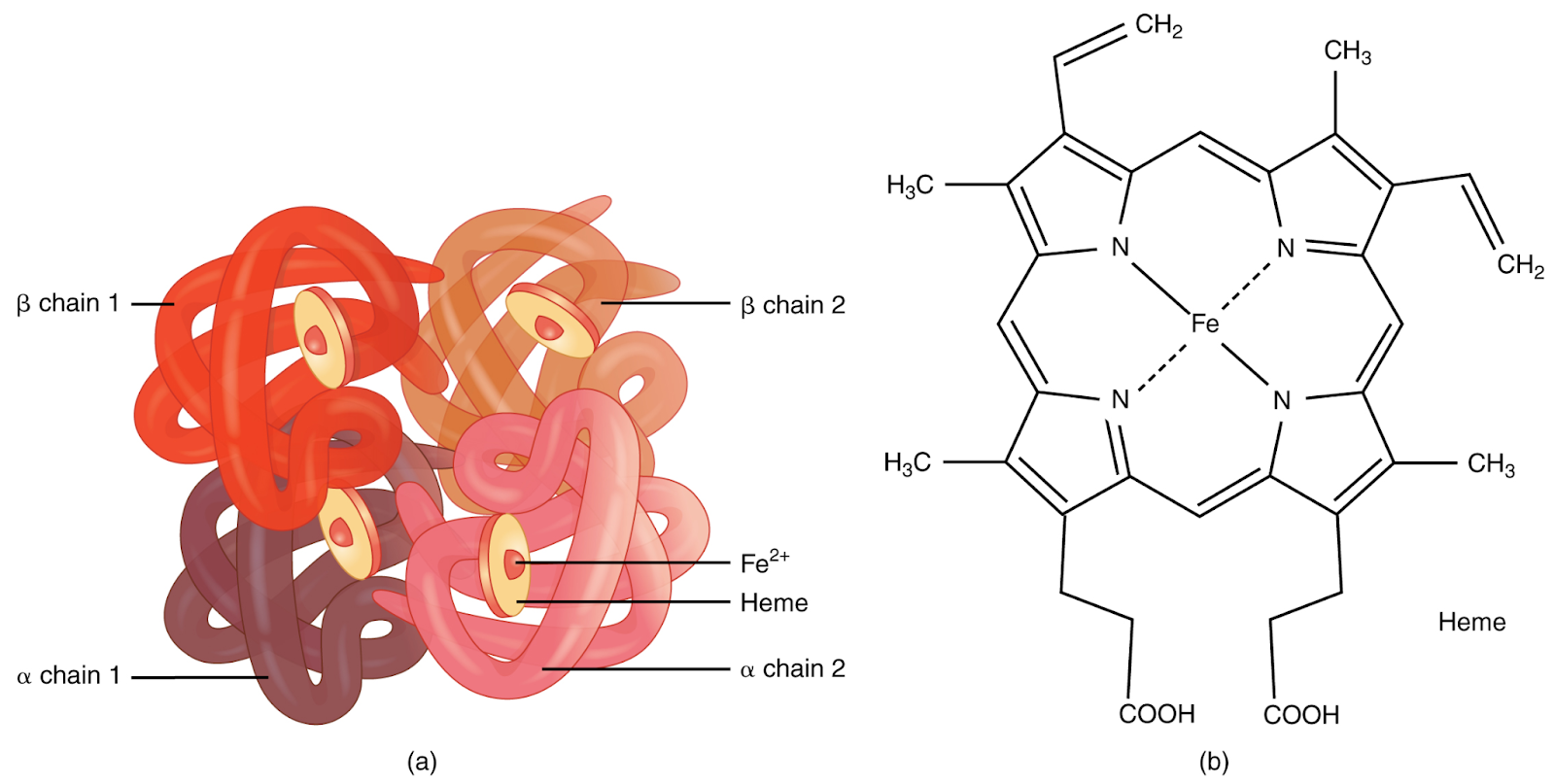
Image courtesy of CNX OpenStax
Interplay of Structures in Protein Function
The functionality of a protein is intrinsically linked to its structure at all four levels. Changes or mutations in any of these levels can have profound effects on the protein's functionality.
- Primary to Quaternary Connections: The sequence of amino acids in the primary structure determines the types of secondary structures that can form, which in turn influence the overall tertiary structure. The tertiary structure is crucial for the assembly and function of multi-subunit proteins in the quaternary structure.
- Structure-Function Relationship: The specific shape of the protein, governed by its structure, is essential for its function. Enzymes, for example, have active sites precisely shaped to bind their substrates.
Structural Proteins vs. Functional Proteins
Proteins can be broadly classified into two categories: structural proteins, which provide support and shape to cells and tissues, and functional proteins, such as enzymes and hormones, which carry out specific tasks within the cell.
- Structural Proteins: These proteins, such as collagen and keratin, typically have repetitive sequences and often form long fibres or sheets.
- Functional Proteins: These are diverse in structure and include enzymes, transport proteins, and signalling molecules.
Crystal Structure Of The Collagen Protein
Image courtesy of Nevit Dilmen
Protein Structure and Diseases
Misfolding or mutations in proteins can lead to diseases. Misfolded proteins can form aggregates, which are implicated in conditions like Alzheimer's disease and Parkinson's disease. Genetic mutations can lead to structural changes in proteins, causing diseases like cystic fibrosis.
- Protein Aggregation: In diseases like Alzheimer's, proteins like amyloid-beta misfold and aggregate, forming plaques that disrupt brain function.
- Genetic Mutations and Protein Dysfunction: A mutation in the CFTR gene alters the structure of a chloride channel protein, leading to cystic fibrosis.
Conclusion
The four levels of protein structure – primary, secondary, tertiary, and quaternary – are fundamental to understanding how proteins function in biological systems. Each level contributes uniquely to the protein's final shape, stability, and functionality. Disruptions at any level can lead to significant biological consequences, highlighting the delicate balance required for proper protein function. This comprehensive understanding of protein structure is crucial for students delving into the complexities of biology and biomedicine.
FAQ
Hydrophobic interactions are crucial in protein folding, primarily influencing the tertiary structure. These interactions occur between nonpolar side chains of amino acids. In aqueous environments, like in the cytoplasm, nonpolar side chains tend to cluster together, away from water. This clustering drives the folding of the protein into its functional three-dimensional shape, as it minimises the exposure of nonpolar areas to the aqueous environment. Hydrophobic interactions are a major force in the stabilisation of the protein's internal core, helping to maintain its overall structure and functionality by ensuring proper folding and alignment of the polypeptide chain.
The specific shape of an alpha helix or beta-pleated sheet in a protein's secondary structure is determined by the nature of the amino acid sequence and the formation of hydrogen bonds. In an alpha helix, the helical shape is due to hydrogen bonds forming between the carbonyl oxygen of one amino acid and the amide hydrogen of another, approximately four residues away. This pattern results in a right-handed helical structure. In contrast, beta-pleated sheets are formed by hydrogen bonding between stretches of polypeptide chains that lie parallel or antiparallel to each other. The particular sequence of amino acids and their side chains affect how these structures fold and align, influencing the overall stability and shape of the secondary structure.
Proteins generally have a single, stable tertiary structure under physiological conditions, which is essential for their specific functionality. However, some proteins can exhibit more than one tertiary structure, a phenomenon known as protein conformational flexibility. This flexibility allows proteins to adopt multiple functional forms, enabling them to interact with different molecules and perform various functions. For example, prion proteins can exist in a normal, non-infectious form and an infectious form; the latter has a different tertiary structure. These alternate forms are not mutations but rather different foldings of the same amino acid sequence, highlighting the dynamic nature of protein structures.
The quaternary structure can greatly influence the function of enzymes, especially those requiring multiple subunits to be fully functional. In enzymes with quaternary structures, the spatial arrangement of the subunits can affect the enzyme's ability to bind substrates, undergo conformational changes necessary for catalysis, and interact with regulatory molecules. Each subunit in a multi-subunit enzyme may have an active site, and the interaction between these subunits can enhance substrate binding and catalytic efficiency, a phenomenon known as allosteric regulation. Additionally, the quaternary structure allows for cooperative binding in some enzymes, where the binding of a substrate to one subunit increases the affinity of other subunits for their substrates. This cooperation is crucial in enzymes like hemoglobin, where it facilitates efficient oxygen transport.
Disulfide bridges significantly contribute to the stability of a protein's tertiary structure. They are strong covalent bonds formed between the sulfur atoms of two cysteine amino acids. When a protein folds, these cysteine residues come into proximity, and an oxidation reaction links them, forming a disulfide bridge. This linkage creates a rigid and stable connection within the protein, anchoring its three-dimensional shape. Such bridges are particularly important in proteins exposed to harsh environments, like extracellular proteins, where they maintain the protein's structure and resist denaturation. In enzymes, they can also maintain the active site's shape, essential for catalytic activity.
Practice Questions
In the secondary structure of proteins, hydrogen bonds play a pivotal role in stabilising the formations of alpha helices and beta-pleated sheets. In alpha helices, hydrogen bonds form between the carbonyl oxygen of one amino acid and the amide hydrogen of another, typically four residues down the chain. This pattern of bonding creates a right-handed helical structure. For beta-pleated sheets, hydrogen bonds form between the carbonyl oxygen of one amino acid and the amide hydrogen of another, but these amino acids are part of separate, adjacent polypeptide chains or different regions of the same chain. These bonds stabilise the sheet-like arrangement of beta strands, which can be parallel or antiparallel.
A mutation in the primary structure of a protein, which involves a change in its amino acid sequence, can significantly affect the tertiary structure and functionality of the protein. Altering even a single amino acid can disrupt the intricate pattern of hydrophobic and hydrophilic interactions, hydrogen bonds, ionic bonds, and disulfide bridges that determine the protein's three-dimensional shape. For instance, in sickle cell anaemia, the substitution of glutamic acid with valine in the haemoglobin protein alters its tertiary structure, leading to the aggregation of haemoglobin molecules and the characteristic sickle shape of red blood cells. This change impairs the ability of haemoglobin to carry oxygen efficiently, demonstrating the profound impact of primary structure mutations on protein function.
